The Hidden Link Between Your Spine and Knee Tendonitis: A Physiotherapist's Guide (ft. Jaeger O'Meara)
Knee tendonitis can be a persistent and frustrating condition that derails athletic careers and daily activities. The road to recovery is often paved with ups and downs, false starts, and frustrations that can leave patients feeling hopeless.
As Physiotherapists, we do well to diagnose and treat the local symptoms of knee tendonitis, but I've increasingly felt we might be missing a crucial piece of the puzzle. Through years of clinical practice, I've observed a surprising pattern: the lower back may play a significant role in the onset, persistence, and recurrence of patellar tendon issues.
In this article, I'll share what I've discovered about this connection and use the case of Australian Football League (AFL) star Jaeger O'Meara to illustrate how spinal dysfunction can manifest as debilitating knee pain.
Key Clinical Insight
In my practice, every patient with knee tendonitis over the past 10-15 years has presented with stiffness in and around the T10-L3 spinal segments - the area between the base of the ribcage and top of the lumbar spine. Addressing this spinal stiffness consistently improves both pain and function.
Understanding Knee Tendonitis (Patellar Tendinopathy)
Knee tendonitis, properly termed patellar tendinopathy, involves dysfunction of the tendon connecting your kneecap (patella) to your shin bone (tibia). Despite the common name "tendonitis," which implies inflammation, most chronic tendon issues actually show minimal inflammatory cells and are better classified as tendinopathies - degenerative changes in the tendon's structure.
This condition is particularly common in jumping sports, earning it the nickname "jumper's knee," but it can affect anyone who repeatedly loads their knee joint.

Common Symptoms of Knee Tendonitis
- Pain and tenderness at the front of the knee, typically at the base of the kneecap
- Thickening of the patellar tendon
- Pain with running, jumping, and hopping activities
- Pain that may lessen during activity but increases afterwards
- In advanced cases, pain that limits athletic performance
- Minimal swelling due to the degenerative nature of tendinopathy
Beyond Overuse: The Real Causes of Tendon Dysfunction
While patellar tendinopathy is often classified as an overuse injury triggered by activities like running, jumping, increased training load, or hard surfaces, this explanation falls short. Tendon loading is normal - our bodies are designed to run and jump. The problem isn't simply that we use our tendons, but how we load them.
Hidden mechanical factors often set the stage for tendon failure when exposed to these external triggers:
- Quadriceps, hamstring, and calf tightness
- Muscle strength imbalances
- Weak gluteal muscles
- Poor core stability
Despite addressing these factors, I continued to see persistent cases until I began looking higher up the kinetic chain - at the spine.
The Spinal Connection: What I'm Finding Clinically
Over the past few decades, I've observed that every patient with knee tendonitis presents with stiffness in and around the T10-L3 spinal segments. This area, comprising the junction between the base of the ribcage and the top of the lumbar spine, shows specific stiffness that patients are often completely unaware of, as it doesn't typically cause back pain.

Why the T10-L3 Area Matters for Knee Health
This specific region of the spine is critically important for two key reasons:
- Femoral Nerve Origin: The T12-L3 segments house the nerve roots that form the femoral nerve, which supplies the quadriceps muscles and courses down the front of the thigh to the knee.
- Dermatomal and Myotomal Connections: These spinal segments correspond to the knee's sensory (dermatomal) and motor (myotomal) connections established during embryonic development.
When these spinal segments become stiff, they can short-circuit normal function of the knee - whether it be increased neural tension, or promoting muscle tightness that acts as a "hand-brake" on the knee, altering how forces are distributed through the patellar tendon during movement.
Case Study: Jaeger O'Meara's Knee Struggle
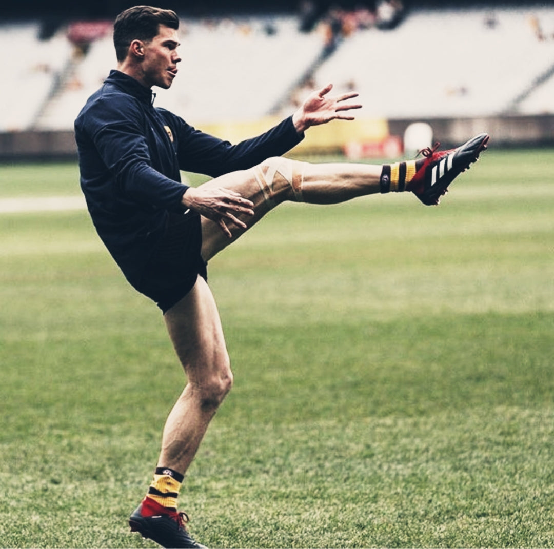
Australian Rules Football star Jaeger O'Meara's career provides a compelling illustration of this connection. After a spectacular start to his AFL career that earned him the 2013 Rising Star Award, O'Meara's career was derailed by persistent knee issues.
Significantly, before his devastating patellar tendon rupture in 2015, O'Meara had already undergone off-season surgery for severe bilateral knee tendonitis that limited his training. While his medical team described the rupture as a "freakish accident," the presence of pre-existing tendon pathology suggests underlying biomechanical issues.
The Spinal Component in O'Meara's Case
Visual analysis of O'Meara's movement and sitting patterns reveals what I term a "spinal hinge" - a specific focal point of bending in the T10-L3 region rather than the distributed curvature we'd ideally like to see. This movement pattern creates excessive stress on specific spinal segments, potentially leading to the stiffness I consistently find in tendonitis patients.

These patterns matter because in moments of high stimulus or fatigue, the body defaults to its most familiar movement pathways. If poor spinal positioning during daily activities (sitting, driving, etc.) creates stiffness, this dysfunction will manifest during athletic performance when demands are highest.
Interestingly, around the same time, the NFL's own Victor Cruz was also dealing with his own patellar tendon rupture. And, in the image above, he also demonstrates the same tendencies through the same parts of his spine.
Comprehensive Treatment Approach
While addressing the spinal component is crucial, effective patellar tendinopathy treatment requires a multifaceted approach:
Conventional Rehabilitation Foundation
- Activity Modification: Finding the balance between sufficient loading for tendon remodelling and avoiding exacerbation of symptoms
- Pain Management: Judicious use of medication for severe pain, with awareness that ice may impede healing by going against what the body is trying to do
- Local Tissue Treatment: Addressing quadriceps flexibility and strength (particularly eccentric strengthening)
- Supportive Measures: Temporary use of taping or bracing to distribute load while underlying issues are addressed

The Missing Piece: Spinal Integration
Based on my clinical findings, I recommend adding these spinal-focused interventions:
- Reduce Spinal Stiffness: Have a Physiotherapist mobilise in and around the T10-L3 segments, or use a lacrosse ball or foam roller for self-mobilisation
- Improve Trunk Strength: Develop comprehensive core stability beyond basic exercises
- Practice Better Spinal Posture: Retrain movement patterns to maintain neutral spinal alignment during daily activities
Conclusion: A New Perspective on Knee Tendonitis
We excel at managing the local symptoms of knee tendonitis, but by incorporating assessment and treatment of spinal dysfunction - particularly around the T10-L3 segments - we may achieve better outcomes for this challenging condition.
The case of Jaeger O'Meara, who eventually returned to play consistent football after his devastating injury, suggests that addressing these underlying biomechanical issues can make the difference between recurrent struggles and sustainable recovery.
If you're struggling with persistent knee tendonitis, I encourage you to discuss spinal assessment with your Physiotherapist. Sometimes the key to resolving stubborn knee pain lies not in the knee itself, but in the intricate connections between your spine and extremities.
Need Personalised Guidance?
If you'd like help trying to uncover the underlying cause of your pain or dysfunction, consider booking an online Telehealth consultation with Grant here!

